Figure 4.
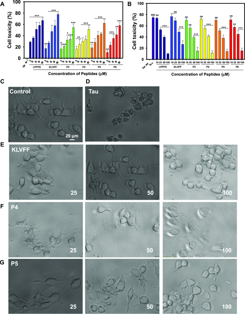
Evaluation of tau-induced cytotoxicity in the presence of different concentrations of peptides. (A) Cell viability of SH-SY5Y cells in the presence of peptides. Cells were incubated with different concentrations of peptides; LPFFD, KLVFF, P3, P4, P5, P6 and (10–100 μM) for 12 h and the viability was measured by MTT assay. DMEM media was used to dissolve the indicated peptides, used as untreated control and set to 100% viability. (B) Full-length tau aggregates (hTau40wt) at 5 μM concentration in the absence or presence of indicated concentrations of peptides (LPFFD, KLVFF, and P3–P6) was added to the SH-SY5Y cells and incubated for 12 h at 37 °C. The cell toxicity was measured by MTT assay. (C–G) Cell imaging. Images of the cells post incubation for 12 h with peptides at various concentrations in the absence and presence of 5 μM tau. The cells were observed under a bright field microscope with a magnification of 20×.

