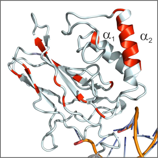
Figure 3.

α2 helix is the most stable secondary structure within the p50-NTD. The p50 residues with ΔGHX greater than 5.5 kcal mol-1 is mapped onto the p50 structure (NTD from PDB id: 1NFK)11 and colored red. The two helices are labeled on the structure.
