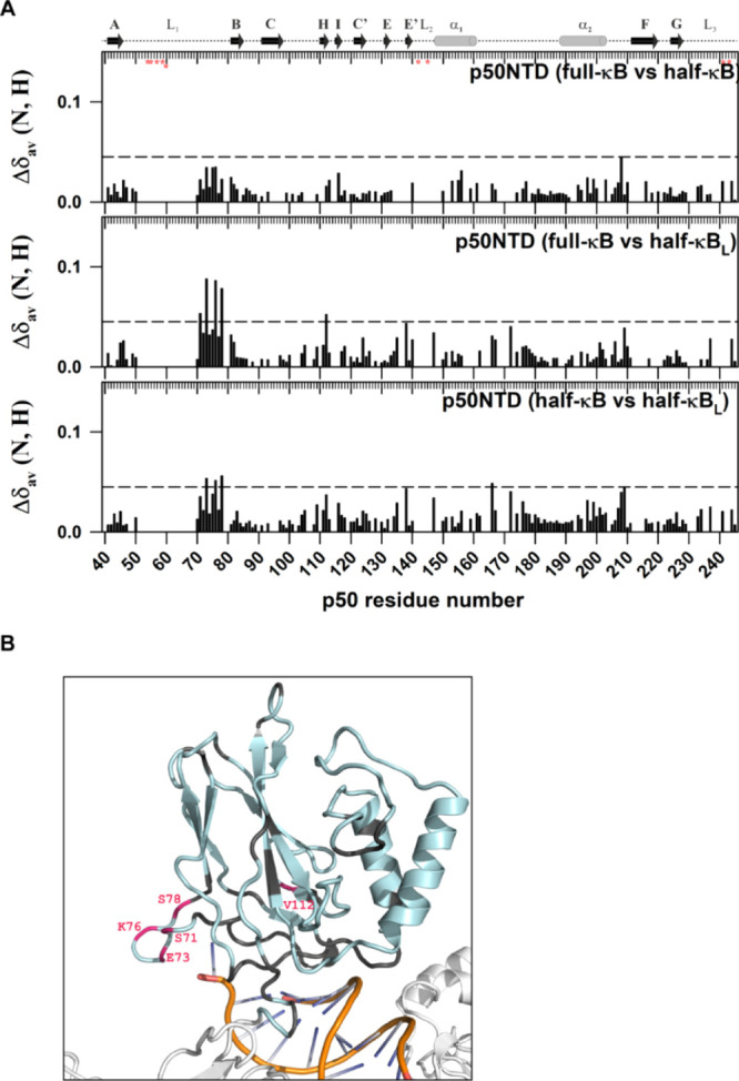
Figure 6.

C-terminal part of the L1 DNA-binding loop interacts with the nucleotides beyond the κB sequence. (A) Plot of CSP observed for the p50-NTD in complex with the full-κB with respect to that with the half-κB (top panel); in complex with the full-κB with respect to that with the half-κBL (middle panel); and in complex with the half-κB with respect to that with the half-κBL (lower panel) DNA-bound forms as a function of the p50 residue number. The other details of the figure are the same as mentioned in Figure 5A. (B) Significant CSPs mapped onto the 3D structure of the p50-NTD (PDB id 1NFK) depicted in pink. The p50-NTD is colored in light-blue; DNA in golden-yellow with bases shown in blue; and the other parts of the p50-RHR structure are shown in white.
