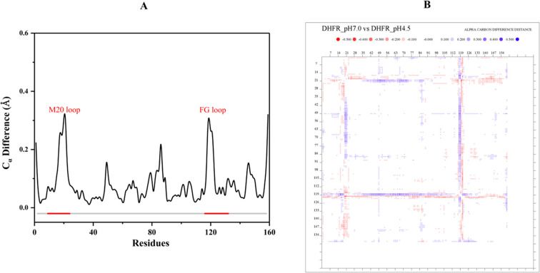
Figure 1.
Structural comparison between the X-ray structures at pH 4.5 and pH 7.0. (A) The Cα difference after superposition shows the most significant changes occur in the Met20 and F–G loop regions. (B) The distance different matrix shows that, in the pH 4.5 structure, the cleft around the folate substrate is slightly closed due to the shifts by the Met20 and F–G loops.

