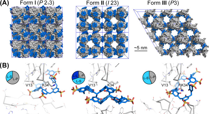
Figure 1.
(A) Crystal packing in RSL-sclx8cocrystal forms I (P213), II (I23), and III (P3). Note the high porosity of the I23 and P3 forms, with nanometer-scale solvent channels. Proteins shown as gray surfaces, and sclx8 shown as blue spheres. (B) Details of the principal protein-sclx8-protein interfaces in each crystal form, with RSL shown as the monomer for clarity. The Val13 and Lys34 binding patch is common to each crystal form. Pie charts show area proportions of sclx8-mediated interfaces (see Table S4). L, ligand; P, protein; S, solvent.

