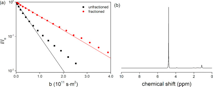
Figure 1.
(a) 1H NMR diffusional decay of the methyl peak in the spectrum of PNIPAM either (■) as obtained or (●) fractioned. The solid line is a single-exponential fit to the initial echo attenuation that provides the average diffusion coefficicent D. (b) 1H NMR spectrum of the solution of the fractioned PNIPAM at 10 mM monomer-equivalent concentration in D2O at 20 °C. The diffusion and electrophoretic NMR experiments were performed on the PNIPAM methyl peak at around 1.1 ppm.

