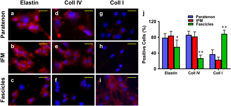
Fig. 12. Immunostaining of isolated cells from paratenon, IFM, and fascicles for expression of structural tendon proteins.
Passage 2 cells were analyzed with ICC. a-c: Elastin; d-f: Collagen IV; g-i: Collagen I; a, d, g: paratenon isolated cells; b, e, h: IFM isolated cells; and c, f, i: Fascicle isolated cells. All components expressed elastin, with high levels in the paratenon and IFM isolated cells (a, b), and minimal levels within fascicle isolated cells (c). High levels of collagen IV staining can be seen in both the paratenon and IFM (d, e), with very few positively stained cells within fascicle isolated cultures (f). Collagen I is apparently expressed at a low level in isolated paratenon (g) and is minimally expressed in IFM (h) cell cultures, while isolated fascicle cells presented high levels of expression for collagen I. (i). Semi-quantification is displayed in figure j, *p < 0.01 (fascicles compared to IFM); **p < 0.001 (fascicles compared to both paratenon and IFM). Yellow bars: 25 μm.

