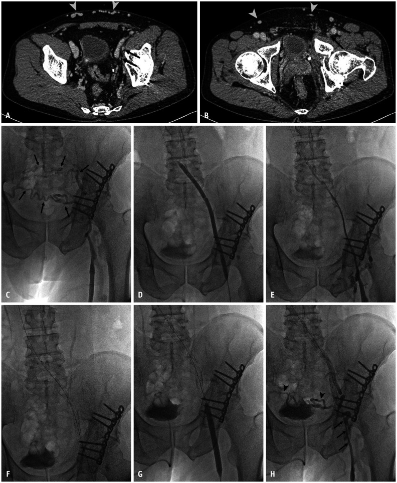
Fig. 4. Endovascular treatment of chronic deep vein thrombosis from left common iliac vein to left femoral vein.
A 60-year-old man presented with left leg swelling and pain in the past year. He had the history of pelvic bone fracture 18 months ago.
A, B. CT shows luminal obliteration of left iliac vein (arrows, A) and left common femoral vein (arrow, B) with multiple dilated collateral vessels along the lower abdominal wall (arrowheads, A, B). C. Venography shows only dilated collateral vessels (arrows) draining into contralateral pelvic vessels, and no deep venous system is observed above the proximal segment of the femoral vein. D. After recanalization of the obliteration segment from the left common iliac vein and femoral vein using a 5-F catheter and a guidewire, sequential balloon angioplasty was performed from left common iliac vein to left femoral vein using a 4 mm × 4-cm balloon catheter (Fortrex; Medtronics) (not shown), and a 7 mm × 20-cm balloon catheter (Mustang; Boston Scientific). E. Follow-up venography shows faint venous flow from the left femoral vein to the left common iliac vein, but this long segmental veins are still collapsed. F, G. Additional stent placement was done for the left common iliac vein and the external iliac vein using two bare metal stents (F), a 12 mm × 10-cm and a 12 mm × 4-cm (E Luminexx; Bard), and balloon angioplasty was done for left common femoral vein and femoral vein using a 8 mm × 8-cm balloon catheter (Conquest; Bard) (G). H. Completion venography shows recovered venous flow from the left femoral vein to inferior vena cava through stents, but the segmental stenosis of the femoral vein and common femoral vein is observed (arrows). Previous noted collateral vessels were reduced but still remain (arrowheads).

