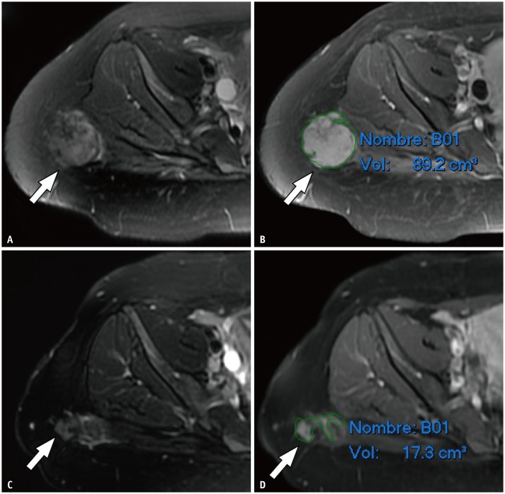
Fig. 1. Magnetic resonance imaging before (A, B) and after (C, D) ablation in a 26-year-old female with desmoid tumor in gluteal region.
A mass with a histological diagnosis of aggressive fibromatosis is observed in the right gluteal region (arrows) on fat-suppressed T2-weighted images (A, C) and contrast-enhanced fat-suppressed T1-weighted images (B, D). Volumetric calculation using a slice-by-slice segmentation allowed the determination of the decrease in tumor volume.

