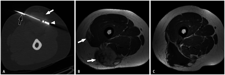
Fig. 2. Ablation technique and outcome in a 33-year-old female with desmoid tumor in the thigh.
A. Computed tomography-guided ablation in prone position. The vertebroplasty needle (black arrow) and microwave probe (arrowhead) are placed inside the tumor (white arrow). B. The axial contrast-enhanced T1-weighted image obtained in supine position before treatment reveals a multifocal desmoid tumor on the posterior lateral aspect of the thigh (arrows). C. Axial contrast-enhanced T1-weighted image obtained in supine position after ablation with microwaves. No enhancement foci that might suggest tumor remnants can be observed at the site of ablation.

