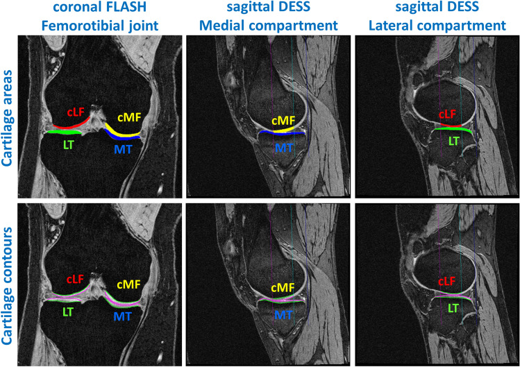
Fig. 1.
Manual segmentation of the femorotibial cartilages [MT/LT medial/lateral tibia, cMF/cLF central (weight-bearing) part of the medial and lateral femoral condyles] from coronal FLASH and sagittal DESS MRI. The figure shows the cartilage areas (top row) and the cartilage contours (bottom; green: total area of subchondral bone; magenta: cartilage surface area). The sagittal MRIs also show the 60% femoral region of interest (magenta line: anterior margin; blue line: posterior end of the condyles; turquoise line: 60% margin)

