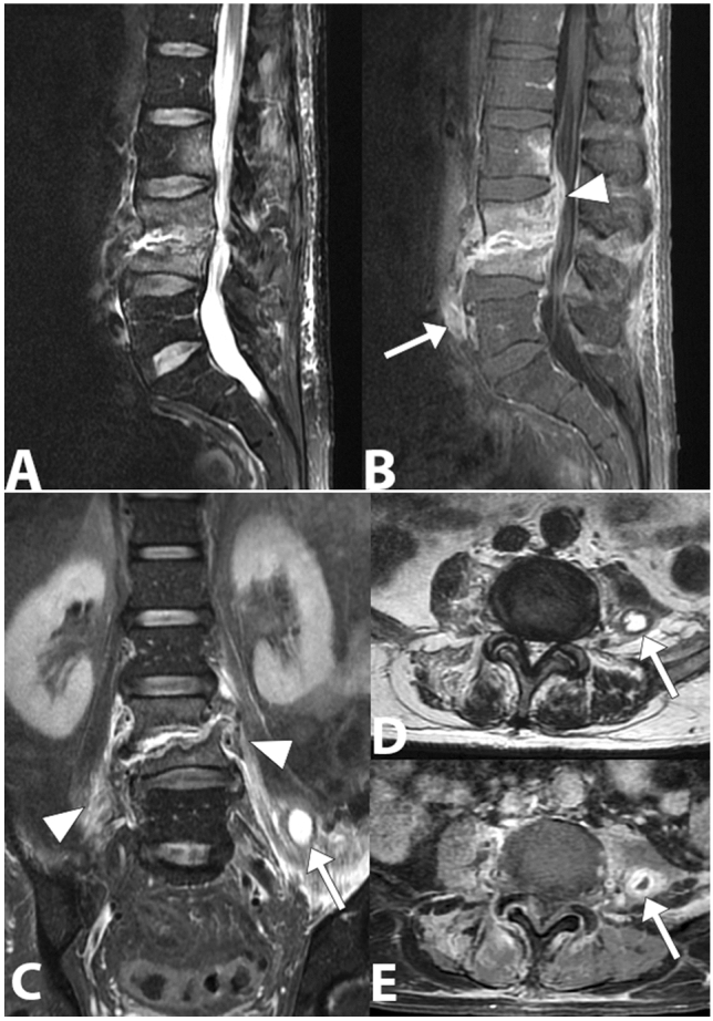
Fig. 10.

Gram-negative (Escherichia coli) spondylodiscitis. Sagittal lumbar spine MRI image (a) showing a high bone marrow signal and partial collapse of the vertebral bodies L3 and L4, destruction of the L3–L4 disc and loss of the definition of the end plate on both sides of the disc. In the T1-weighted sagittal image obtained with saturation of the fat signal and after the injection of the contrast medium (b), a better definition of the involvement of the paravertebral (arrow) and epidural space with narrowing of the vertebral canal (arrowhead) can be observed. The posterior L2 vertebral body is also involved. In coronal STIR image (c), axial T2- weighted (d) and axial T1- weighted with fat saturation and gadolinium (e), edema and inflammatory exudate can be observed in the paravertebral soft tissue (arrowheads) and abscesses in the left psoas muscle (arrow)
