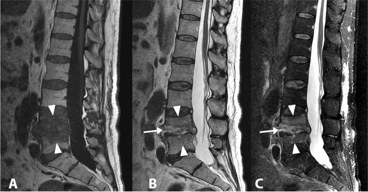
Fig. 6.
Lumbar (L4-L5) spondylodiscitis caused by Staphylococcus aureus. The whole vertebral body of L4 and the upper portion of L5-vertebral body show an altered signal intensity in the T1-weighted image (a), in the T2-weighted image (b) and in the fat saturated T2-weighted image (c) (arrowheads). The L4–L5 disc is involved and it appears thinned, with an increased signal intensity in the T2-weighted and fat saturated T2-weighted images (arrows)

