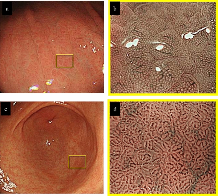
Fig. 2.
Endoscopic findings. a Conventional white-light endoscopic findings of the greater curvature in the middle part of the corpus. b The view of the reddened gastric areas by magnifying endoscopy with narrow-band imaging (ME-NBI). c Conventional white-light endoscopic findings of the gastric antrum. d ME-NBI findings of the pyloric gland mucosa. The microvascular pattern of the fundic gland mucosa showed a dilated SECN with regular honeycomb-like arrangement and collecting venules, and the microsurface pattern depicted a regular oval crypt opening and normal oval MCE (a, b). Both conventional white-light and ME-NBI findings showed that the gastric pyloric gland mucosa was normal (c, d). ME-NBI magnifying endoscopy with narrow-band imaging, SECN subepithelial capillary network, MCE marginal crypt epithelium

