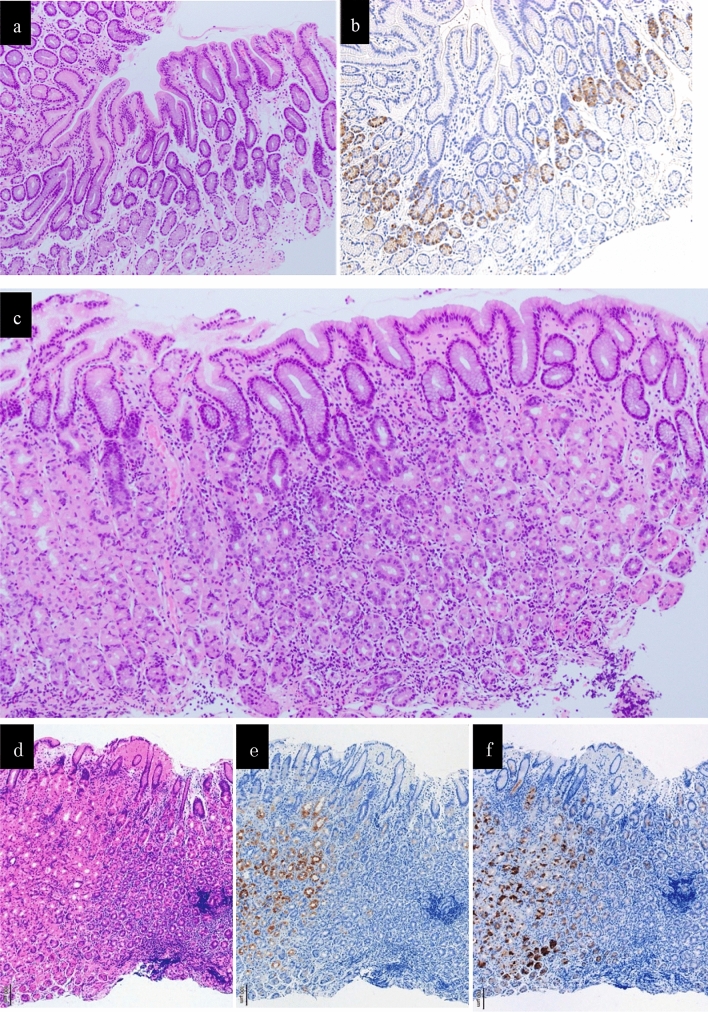
Fig. 3.
The histopathological findings. a The histopathological findings of the specimens biopsied from the lesser curvature in the pyloric gland mucosa did not show either atrophy or intestinal metaplasia (HE staining). b The histopathological findings of the specimens biopsied from the lesser curvature in the pyloric gland mucosa showed mild hyperplasia of G-cells (immnostaining for gastrin). c The histopathological findings a biopsy specimen obtained from the greater curvature in the middle part of the gastric corpus did not show atrophy, intestinal metaplasia, or decreased parietal cells accompanied with a dense lymphocyte infiltration without neutrophils in the middle-to-deep part of the lamina propria mucosa (HE staining). d The finding of HE staining of a biopsy specimen obtained from the greater curvature in the middle part of the gastric corpus. e The finding of immunostaining for H +/ K+ -ATPase of a biopsy specimen obtained from the greater curvature in the middle part of the gastric corpus. f The finding of immunostaining for Pepsinogen I of a biopsy specimen obtained from the greater curvature in the middle part of the gastric corpus. The immunostaining for both H+/K+-ATPase and Pepsinogen I was negative in the area with a dense lymphocyte infiltration (d, e, f)

