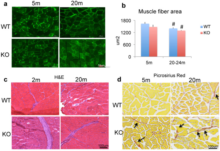
Figure 4.
Histomorphometric analysis of muscle from WT and Fgf2KO mice. Muscle sections from tibialis anterior muscle in 5-month and 20–24 months old WT and Fgf2KO mice. (a) Laminin-stained sections reveal overall reduced myofiber cross sectional area is observed within old WT and old Fgf2KO mice. (b) Quantitative analysis of cross-sectional area of skeletal muscle from 3 mice/group revealed significant reduction in old WT and old Fgf2KO compared with respective young genotype. #Compared to corresponding 5 m p < 0.05. (c) Muscle sections from tibialis anterior muscle in young and old (2 months and 22 months) WT and Fgf2KO stained with hematoxylin and eosin reveal large swaths of ECM/collagen deposition and increased inflammatory infiltrate in young and old Fgf2KO as well as old WT. (d) Picroriues red staining for collagen showed increased fibrosis (arrows) in the muscle of 20 m-WT and 5 m Fgf2KO compared with 5 m WT, and further increased in 20 m Fgf2KO.

