Figure 3.
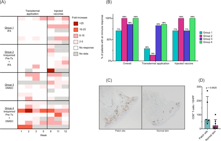
CD8+ immune responses to class I MHC-restricted melanoma peptides (12MP) in in vitro stimulated (IVS) ELIspot assay. (A) Heatmap demonstrating the magnitude of immune response to transdermal and injected vaccines. Each patient is represented as a single row. Observed immune responses with fold increase over negative control less than 2 x or those not meeting other criteria for response are shown in white. Gray represents unavailable data. (B) Percent of patients with an immune response to 12MP by group in the transdermal and injected time points. (C) Immunohistochemistry of a patch site biopsy and adjacent normal skin from one patient (group 4) stained with CD8 antibody demonstrating dense infiltrate of CD8+ cells compared with the normal skin. (D) Comparison of overall CD8+ T cell counts in patients from group four patch sites versus normal skin (median, IQR), with a trend toward higher CD8+ T cell count in the patch site; p=0.0625. Comparison was made using the Wilcoxon rank sum test with alpha=0.05. DMSO dimethylsulfoxide; HPF, high power field; IFA, incomplete Freund’s adjuvant; Tx, treatment

