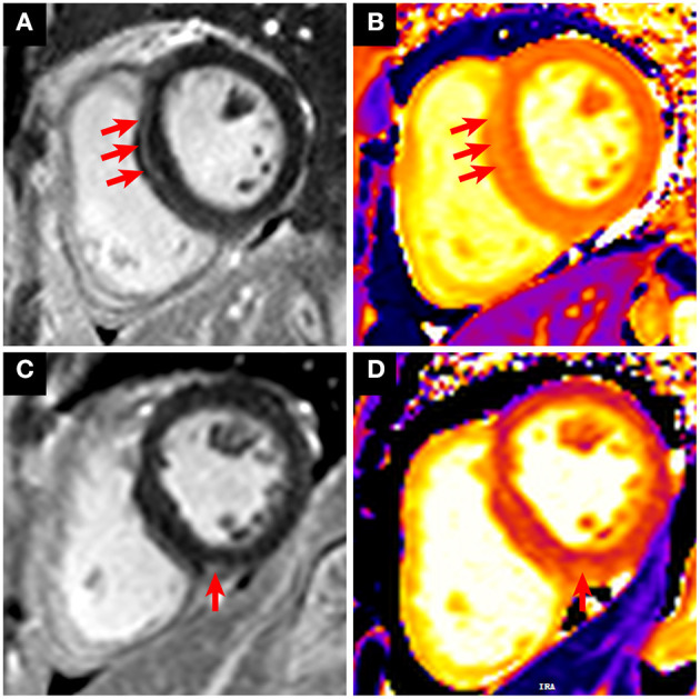
Figure 2.

Representative CMR images with LGE positive in patients who recovered from COVID-19. (A,B) A 31-year-old male patient with cardiac injury underwent CMR 6 months after recovery from COVID-19. The short-axis LGE sequence showed enhancement in the LV septal segment (A, red arrows). Increased native T1 was shown in the corresponding location of focal LGE (B, red arrows). (C,D) A 63-year-old female patient with cardiac injury underwent CMR 6 months after recovery from COVID-19. The short-axis LGE sequence showed enhancement in the right ventricular insertion point (C, red arrow). Increased native T1 was shown in the corresponding location of focal LGE (D, red arrow).
