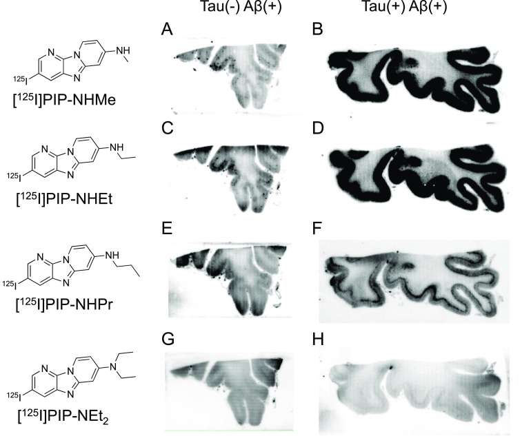
Figure 3.
Comparison of in vitro autoradiography of (A, B) [125I]30 ([125I]PIP-NHMe), (C, D) [125I]31 ([125I]PIP-NHEt), (E, F) [125I]32 ([125I]PIP-NHPr), and (G, H) [125I]40 ([125I]PIP-NEt2) in brain sections from an AD patient. A, C, E, and G show results in Aβ(+)/tau(−) brain sections. B, D, F, and H show results in Aβ(+)/tau(+) brain sections.

