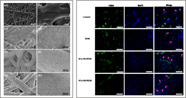
Figure 2.
Left: SEM images showing the morphology of substrates with different amount of CuNPs at two magnifications: (a, b) outer eggshell membrane (ESM), (c, d) 0Cu-BG/ESM, (e, f) 2Cu-BG/ESM, and (g, h) 5Cu-BG/ESM. Right: Detection of increased vessel by immunofluorescence of CD31 (green) at day 7. Nuclei are stained with DAPI (blue). Vascularized areas are indicated by pink arrows. Scale bar = 100 μm. Reprinted with permission from ref (68). Copyright 2016 Elsevier.

