Figure 5.
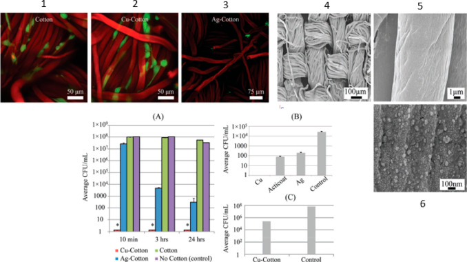
Live–dead staining at laser scanning confocal microscopy are in picture 1, 2, and 3 (red cells = dead, green cells = dead, fibers = red for autofluorescence). SEMs of copper–cotton substrates are in picture 4, 5, and 6. Graph A: Antimicrobial activity against A. baumannii at different times. Graph B: A direct comparison among Cu- and Ag-coated cotton substrates and a commercial silver wound dressing, Acticoat. Graph C: Plot showing about 3-log kill for Cu-cotton samples in the presence of A. baumannii. Reprinted with permission from ref (77). Copyright 2011 John Wiley and Sons.

