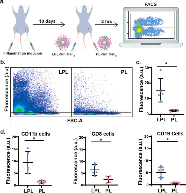
Figure 4.
Immune targeting of inflamed lymph nodes in vivo. (a) Schematic illustration of the in vivo experimental setup used for the injection of both PL-Sm:CaF2 (nanofluorides, i.e., PL) or LPL-Sm:CaF2 (glyconanofluorides, i.e., LPL) NCs into inflamed mice footpads (20 μL of 25 mg/mL NCs). (b) Representative dot blots of FACS analysis of cells excised from lymph nodes 2 h post-injection of LPL-Sm:CaF2 or PL-Sm:CaF2 NCs. (c) Quantitative analysis of the FACS data (rhodamine) obtained from five different mice (N = 5, Student’s test, * represents a p value <0.05). (d) Dot graph representing the lymphatic distribution of LPL-Sm:CaF2vs PL-Sm:CaF2 within subtypes of immune cells that were excised from lymph nodes 2 h post-injection of the NCs (N = 5), from left to right: CD11b leukocyte cells, CD8 T-cells, and CD19 B-cells. All studies were performed with fluorescently labeled nanofluorides (either LPL-Sm:CaF2 or PL-Sm:CaF2).

