
Figure 5.
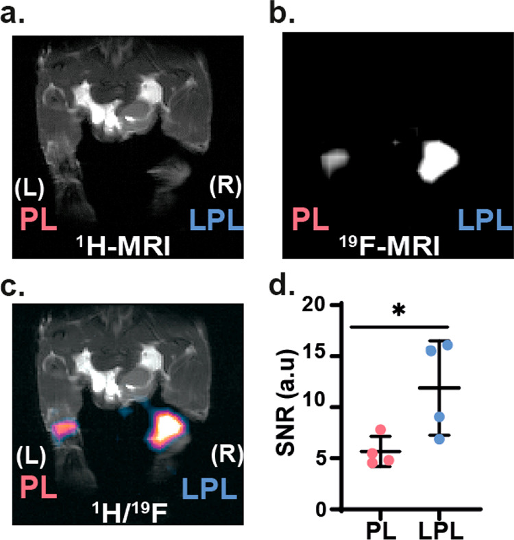
In vivo19F MRI study of inflamed mice. Ten days post-immunization, mice (N = 4) were subcutaneously injected with LPL-Sm:CaF2 NCs (right leg, labeled as “R”) and PL-Sm:CaF2 NCs (left, labeled as “L”). Then, 2 h post-injection mice were anesthetized and scanned with MRI. (a) 1H MRI, (b) 19F MRI, and (c) 1H/19F MRI overlay of a representative mouse. (d) SNR of 19F MRI at the LNs ROIs (N = 4, Student’s test, * represents a p value <0.05). The in-plane resolutions of the 1H MR and 19F MR images are 0.35 × 0.2 mm2 and 1.4 × 0.78 mm2, respectively; the slice thickness is 1 mm in 1H MRI and 0.78 mm in 19F MRI.