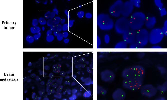
Figure 3.
MMNG HOS transforming gene (MET) amplification test by fluorescence in situ hybridization (FISH). MET is represented by a single red dot, and centromere probe of chromosome 7 (CEP7) is represented by a single green dot. Amplification test by FISH revealed no MET amplification (MET/CEP7 ratio <2.0 and MET per cell<5) in the primary lung tumor per-crizotinib but a cluster MET amplification (MET/CEP7 ratio ≥2.0 and MET per cell≥5) in the metastatic brain tumor post-crizotinib.

