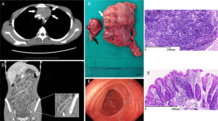
Figure 1.
Diagnostic Evidence of Thymoma with Autoimmune Enteropathy and Myocarditis. An axial, CT contrast-enhanced scan of the chest (A) shows a mediastinal mass (arrows) invading into the left anonymous vein. During surgery, we found that not only the left anonymous vein (white arrow) but also the anterior segment of left-upper lobe (black arrow) were involved by the thymoma (B). The pathological sections of tumor stained with hematoxylin and eosin shows typical B1 thymoma (C). A coronal, CT contrast-enhanced scan of the abdomen and pelvis (D) shows the typical thickening and high-density of the colon wall. The enteroscopy (E) showed the sigmoid colonic mucosa was markedly thinner than normal, and the colon mucosal biopsies (F) detected the colonic mucosa with chronic inflammation, mild lymphocytosis, and extensive loss of goblet cells.

