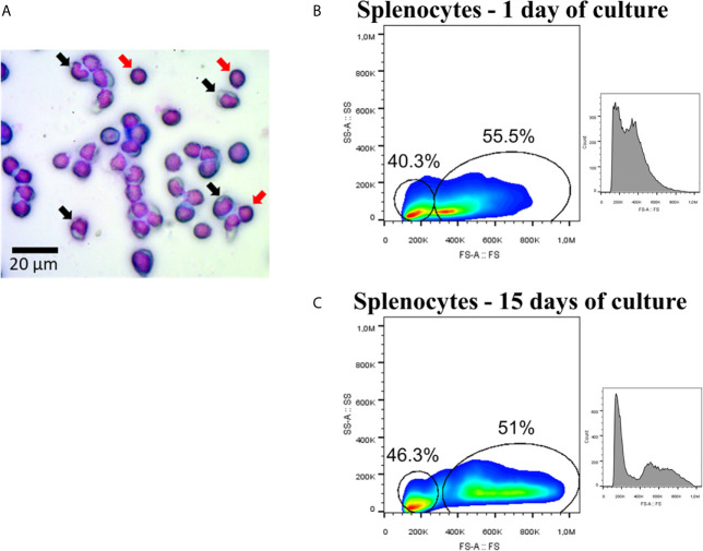
Figure 1.
Splenocytes of Atlantic salmon. (A) Mononuclear fraction. Black arrows showed cells with morphology associated with monocytes, while red arrows showed examples of cells with lymphocyte-associated morphology. The image was taken with magnification 400×. (B) Flow cytometry (FS-A/SSC-A and histogram/FS-A) using splenocytes at 1 day of culture. (C) Flow cytometry (FS-A/SSC-A and histogram/FS-A) using splenocytes at 15 day of culture.

