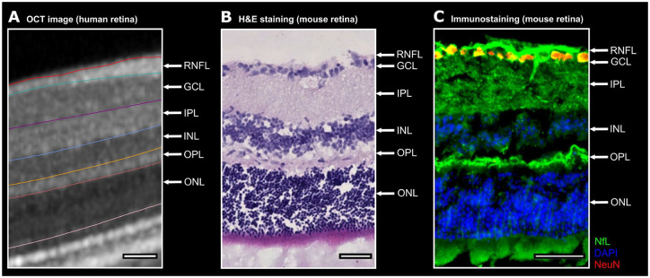
Figure 2.
NfL and its expression pattern within the intra-retinal layers. A. One representative human OCT image with the boundaries between the intra-retinal layers is shown. B. The retinal OCT image is juxtaposed to a C57BL/6J mouse retina. The mouse retina is depicted by H&E staining. C. One representative retinal cross-section from a mouse unveils a predominant expression of neurofilament (green) in the RNFL, GCIPL and OPL. The nuclear and cytosolic antigen NeuN (red) is commonly used to identify neurons. For a better separation of the individual intra-retinal layers the nuclei were counterstained with DAPI (blue) a fluorescent dye strongly binding to DNA.
Scale bars 50 µm. DAPI, 4′,6-Diamidin-2-phenylindol; GCIPL, ganglion cell and inner plexiform layer; H&E, hematoxylin and eosin; NeuN, neuronal nuclear antigen; NfL, neurofilament light chain; OCT, optical coherence tomography; OPL, outer plexiform layer.

