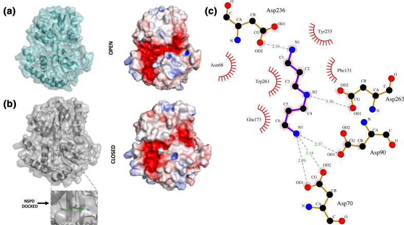
Fig. 5.
Homology models of NspS in open and closed form with docked norspermidine. (a) Homology model of the NspS open conformation with ribbon and transparent surface is shown in teal. The electrostatic surface representation of the open conformation was calculated in Pymol. Red shows negatively charged regions, blue are positive and white is neutral. (b) Homology model of the NspS closed conformation with ribbon and transparent surface in grey. Norspermidine (green sticks) is docked into the model. The electrostatic surface representation of the closed conformation is shown with same colouring as (a). (c) LigPlot representation of norspermidine docked to the closed conformation of the NspS homology model. H-bonding interactions between norspermidine (blue sticks) and residues D70, D90, D236 and D263 are shown with dashed grey lines. Length of H-bonds are shown with green numbers. Residues that form hydrophobic interactions with norspermidine are shown with red eyelashes. Figures were created using Pymol and LigPlot.

