Figure 4.
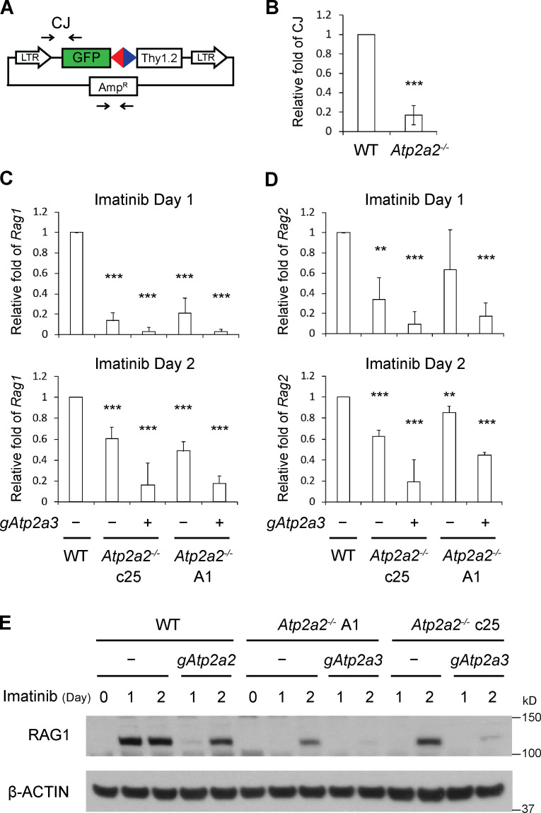
Loss of SERCA2 and SERCA3 leads to diminished Rag expression. (A) Schematic of pMGINV with locations of PCR primers (arrows) used to amplify CJ and the plasmid backbone (AmpR). (B) Quantitative PCR analysis of pMGINV CJ formation in plasmids recovered from WT and Atp2a2−/− abl preB cells treated with imatinib (mean ± SD; n = 3). ***, P < 0.001. (C and D) Levels of Rag1 (C) and Rag2 (D) mRNA assayed by quantitative RT-PCR in abl preB cells treated with imatinib for 1 or 2 d. WT and Atp2a2−/− abl preB clones (−) were assayed, as were Atp2a2−/− abl preB clones with bulk Atp2a3 inactivation (+). Rag1 and Rag2 mRNA levels were normalized to β-actin mRNA and relative to the WT value, which was set at 1. Rag1 and Rag2 mRNAs were not detected at significant levels in cells not treated with imatinib (mean ± SD; n = 3). **, P < 0.01; ***, P < 0.001 compared with WT. (E) Western blot analysis for RAG1 and actin in WT and Atp2a2−/− abl preB cell clones (−) and cells bulk Atp2a2 (gAtp2a2) or Atp2a3 (gAtp2a3) inactivated and treated with imatinib for 1 or 2 d (n = 1 for 1 d, n = 2 for 2 d).

