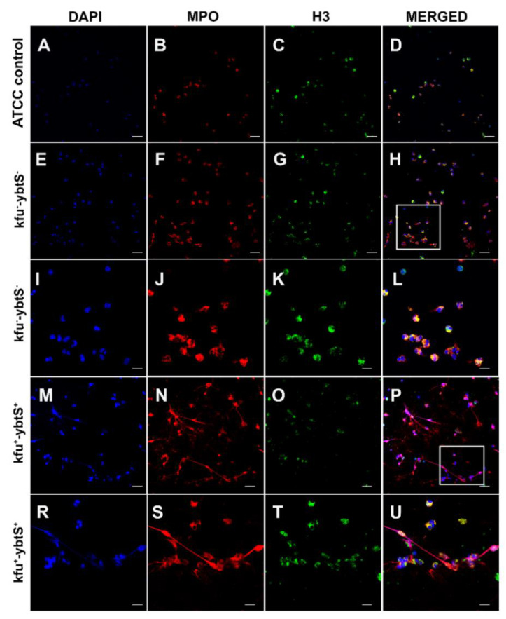
Figure 2.
Confocal microscopic images of NETs. The samples were stained consecutively with myeloperoxidase (MPO, red) and histone 3 (H3, green). The nuclei were counterstained with DAPI (blue). Neutrophils were seen intact with K. pneumonia ATCC 700,831 control (A–D). The kfu- -ybtS- isolates depicted rare and weak NET formation (E–L). The rectangular area in image H was magnified in images I–L. The kfu+ -ybtS+ isolates showed abundant NET formation with excessive histone and MPO release in extracellular matrix (M–U). The rectangular area in image p was magnified in images R–U. Bars: A–H, M-p = 25 μm; I–L, R–U = 10 μm.

