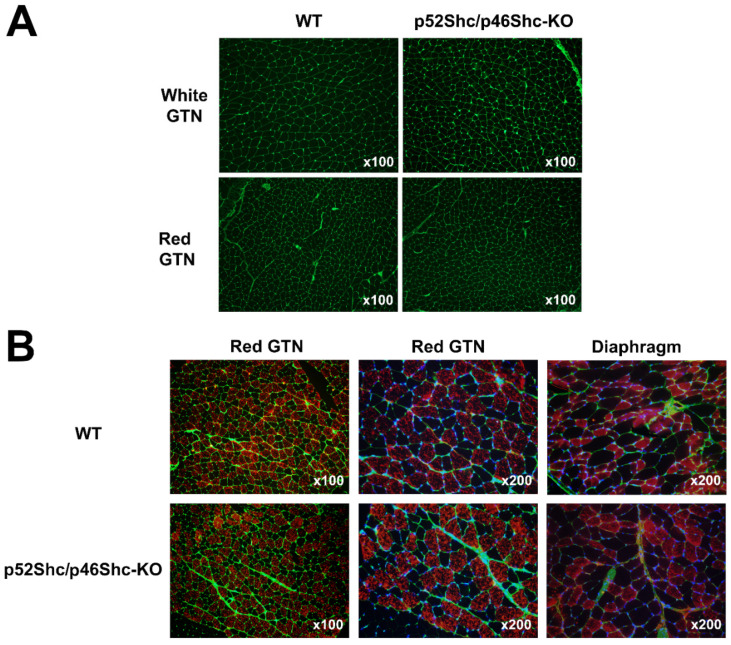
Figure 4.
Comparison of GTN muscle and diaphragm morphology and fiber type composition of p52Shc/p46Shc-KO rats and WT littermates. (A) Cross-sections of white (glycolytic) and red (oxidative) areas of GTN muscle stained with WGA-fluorescein (green staining) are presented. (B) Cross-sections of red (oxidative) areas of GTN muscle and diaphragm stained with anti-slow MHC antibody (red staining) and WGA-fluorescein (green staining) are presented. DAPI (blue staining) was used to visualize nuclei.

