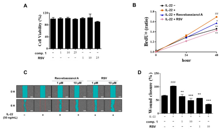
Figure 4.
Effects of resvebassianol A on the proliferation and migration of HaCaT cells. (A) Cells were cultured in 96-well plates, and they were treated with resvebassianol A and RSV (1, 10, and 25 μM). After 24 h cell viability was measured using the MTT assay. (B) HaCaT cell proliferation after 24 and 48 h of treatment with resvebassianol A and RSV was measured using BrdU incorporation assay. (C) The wound margin was photographed after 0 h and 6 h of wound scratching. (D) Quantitative analysis of wound closure was determined as the wound area at a given time relative to that of the IL-22-treated group. Values are expressed as means ± SEM. ## p < 0.01 and ### p < 0.001 versus untreated (control) group; * p < 0.05, ** p < 0.01, and *** p < 0.001 vs. IL-22-treated group.

