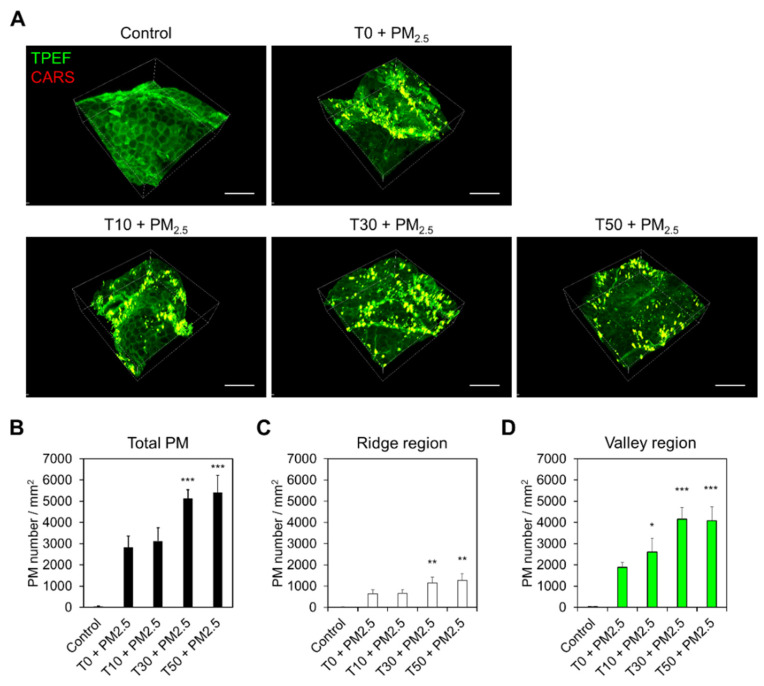
Figure 3.
Visualization of PM2.5 deposition on the skin surface by skin barrier disruption. (A) En face MNLO 3D images of the label-free stratum corneum (TPEF, green) and PM2.5 (CARS, red) in PM2.5-exposed human skin biopsy samples after tape-stripping (T). T10, T30, and T50 indicate strippings of 10, 30, and 50 times, respectively. Scale bars, 100 μm. (B–D) Number of PM2.5 particles on the skin surface, ridge, and valley regions by the number of tape-strippings (mean ± S.E.M., * p < 0.05, ** p < 0.01, *** p < 0.001; two-tailed Student’s t-test).

