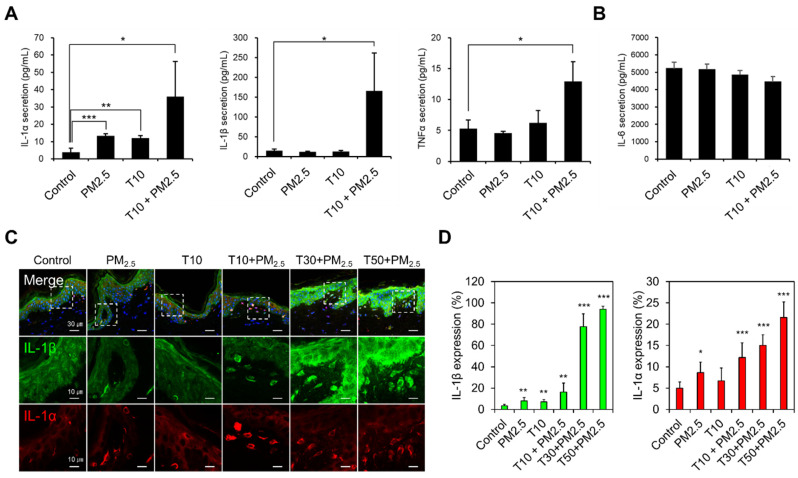
Figure 5.
Inflammatory response induced by PM2.5 exposure and skin barrier disruption. (A,B) Secreted cytokines (IL-1α, IL-1β, TNF-α, and IL-6) measured in culture medium by ELISA. (C) Confocal images of cross-sectioned PM2.5-exposed human skin biopsy samples after tape-stripping (T). IL-1β (green) and IL-1α (red) were stained with antibodies, and nuclei were counter-stained with DAPI (blue). The white dashed boxes in the merged images (upper row) are enlarged in the separated images (lower rows). Scale bars, 30 μm for the merged images and 10 μm for the enlarged images. (D) The cells with a high expression of IL-1β and IL-1α were quantified in each condition (mean ± S.E.M., * p < 0.05, ** p < 0.01, *** p < 0.001; two-tailed Student’s t-test).

