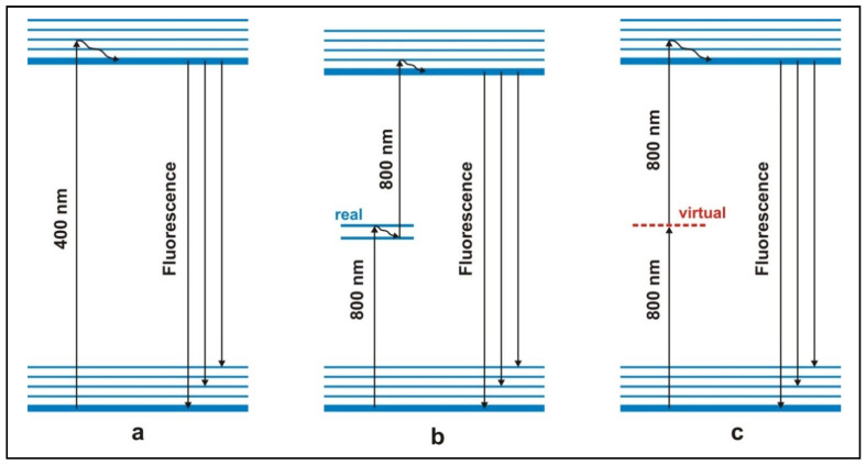
Figure 1.
Different modes of excitation of fluorescence. (a) Left: conventional fluorescence excitation by one photon (e.g., 400 nm). (b) Middle: excitation by stepwise absorption of two photons via a real intermediate energy level (e.g., 800 nm, preferably from a nanosecond laser). PRINCIPLE OF DERMATOFLUOROSCOPY. (c) Right: excitation by simultaneous absorption of two photons via virtual energy level (e.g., 800 nm, preferably from a femtosecond laser). This mode of excitation is used in femtosecond laser spectroscopy. Due to the small cross-section, a nanosecond (ns) pulse excitation with tolerable intensities gives rise to only an extremely weak fluorescence.

