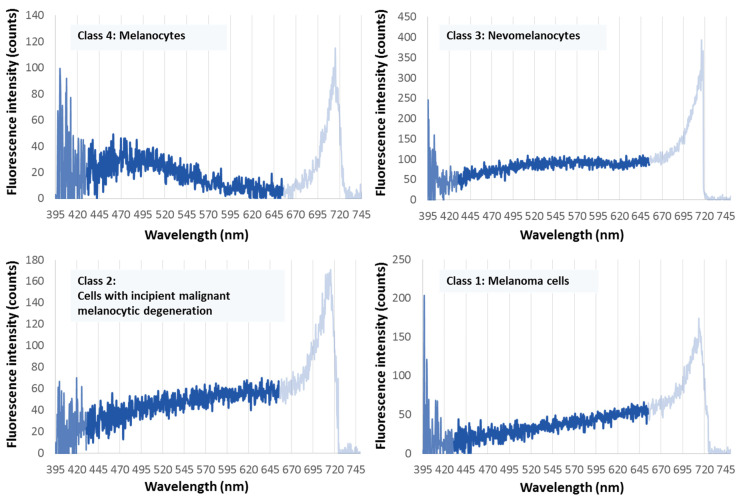
Figure 2.
The four characteristic melanin fluorescence spectra of pigmented skin cells in the spectral range between 430 nm and 650 nm. Upper left: melanocytes; upper right: nevomelanocytes; lower left: dysplastic nevomelanocytes; lower right: melanoma cells. The fluorescence is excited by stepwise two-photon absorption with 800 nm/2 ns pulses (principle of dermatofluoroscopy). (The intensity above 650 nm results from another non-linear process not considered here. In any case, it is ensured that its intensity is zero below 650 nm.)

