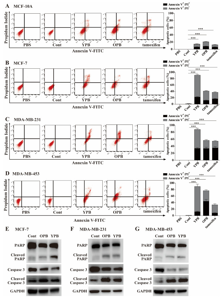
Figure 3.
Evaluation of apoptosis in breast cancer cells treated by the peptides. (A–C) Flow cytometric analyses of breast cancer cells treated by the peptides. MCF-10A (A), MCF-7 (B), MDA-MB-231 (C), and MDA-MB-453 (D) cells were individually treated by PBS, 30 µM of Cont, YPB, and OPB peptides (unlabeled), and 15 µM of tamoxifen for 48 h, followed by the staining of Annexin V-FITC and PI. The apoptotic rates of the treated cells were analyzed by flow cytometry with representative images shown at left and quantitative apoptotic rates calculated by FlowJo software shown at right. Data represent the mean ± S.D. * p < 0.05, ** p < 0.01, *** p < 0.001. (E–G) Western blot analyses of apoptotic markers in breast cancer cells treated by the peptides. MCF-7 (E), MDA-MB-231 (F), and MDA-MB-453 (G) cells were treated as described in (A–C). The cell lysates were subjected to Western blot analyses using indicated antibodies with GAPDH as a loading control.

