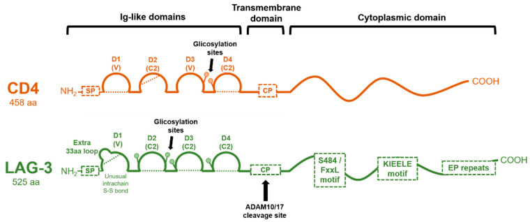
Figure 2.
Molecular organization of CD4 and LAG-3 proteins. The domain organization of CD4 and LAG-3 is schematically shown in the figure, with each Ig-like domain indicated as arcs. Dotted lines represent disulfide bonds. The cleavage site for ADAM 10/17 in LAG-3 is shown, rendering a soluble version. CP: connecting peptide; SP: signal peptide.

