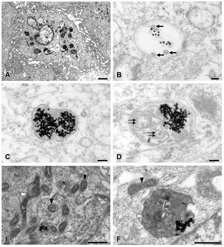
Figure 6.
Transmission electron microscopy of the 30 ppm AuNP-treated pericyte group at low (A) and high magnifications (B–F). Representative images are shown for late endosomes containing AuNPs and multivesicular bodies (B), autolysosomes containing only AuNPs (C), and autolysosomes containing AuNPs and intracellular debris (D). Affected mitochondria showed mild to severe swelling (E,F). Note the AuNPs (electron-dense dots), multivesicular bodies (black arrows), intracellular debris (double black arrows), unaffected mitochondria (black arrowhead), and swollen or damaged mitochondria (white arrowhead). Bars: 1000 nm (A), 100 nm (B–D), 500 nm (E,F).

