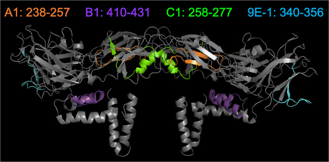
Fig. 4.
Mapping of the binding epitopes of mAbs A1, B1, C1, and 9E-1 on the ZIKV E protein dimer. The binding epitope of mAb A1, B1, C1, or 9E-1 is marked in orange, purple, green, or cyan color on the dimeric form of ZIKV E protein (PDB ID: 5IRE), respectively. The binding epitope of mAb A1 (orange color) is buried underneath two beta-sheets. The binding epitope of mAb C1 (green color) is located in the interface between two ZIKV E subunits. The binding epitope of mAb B1 (purple color) is located near the stem domain of ZIKV E protein. The binding epitope of mAb 9E-1 (cyan color) is located in the domain III of ZIKV E protein

