Figure 1.
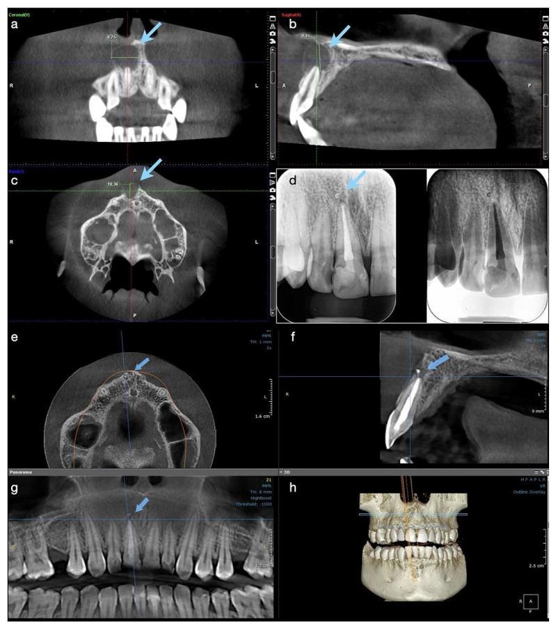
Case 1. Radiological investigations for tooth #11: (a–c) Initial CBCT scans with the measurements of the lesion’s size, indicated by blue arrows: mesiodistal diameter 9.75 mm, height 9.31 mm, buccolingual diameter 10.36 mm, with the interruption of the cortical buccal plate (9.75 × 9.31 × 10.36 mm); (d) control periapical X-ray at 6 months, showing the quality of the endodontic treatment and almost the complete healing of the lesion; (e–g) CBCT scans at 12 months showing the intact buccal cortical plate, the formation of new bone, and the almost complete healing of the lesion. A small enlargement of the periodontal ligament space is still observed apically, (h) 3D reconstruction where intact maxillary bone is observed.

