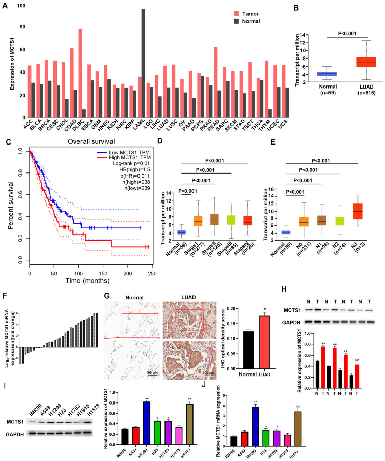
Figure 1.
MCTS1 expression is upregulated in LUAD tissues, and closely associated with prognosis and clinicopathological characteristics. (A) MCTS1 expression in TCGA database and comparisons with other tissues. (B) MCTS1 expression in healthy controls and LUAD tissues was detected based on TCGA database. (C) High MCTS1 expression was associated with unfavorable outcomes in patients with LUAD. MCTS1 was expressed at high levels along the LUAD (D) stages and (E) metastasis. (F) RT-qPCR analysis demonstrated that MCTS1 mRNA expression was upregulated in LUAD tissues. (G) Representative MCTS1 immunostaining in healthy and LUAD samples. (H) Western blot analysis demonstrated that MCTS1 protein expression was upregulated in LUAD tissues compared with normal tissues. (I) Western blot and (J) RT-qPCR analyses were performed to detect MCTS1 protein expression in different LUAD cell line. *P<0.05, **P<0.01 vs. normal or IMR90 cells. MCTS1, multiple copies in T-cell lymphoma-1; LUAD, lung adenocarcinoma; TCGA, The Cancer Genome Atlas; RT-qPCR, reverse transcription-quantitative PCR; IHC, immunohistochemistry; N, normal; T, tumor.

