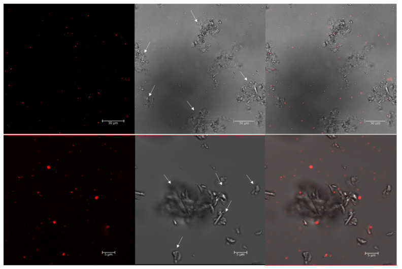
Figure 1.
Confocal images of Nile red-stained Haloferax mediterranei cells. The Hfx. mediterranei cells in the stationary phase were stained with Nile red (0.5 µg/mL) and imaged with a confocal microscope 4 days after subculture. Arrows point to crystals of mineral salts. From left to right: red fluorescence PHA granules; phase contrast image; overlay image of red fluorescence and phase contrast image.

