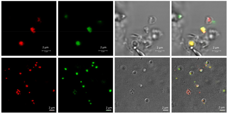
Figure 2.
Fluorescence confocal micrographs of living Haloferax mediterranei cells, with PHA granules stained with Nile red and SYBR Green. Unfixed Hfx. mediterranei cells were co-stained with Nile red lipid fluorescence dye and the nucleic acid-staining dye SYBR Green, revealing the intracellular co-localization of PHA granules with numerous DNA spots in the cytoplasm of the cell, as visualized during confocal microscopy. From left to right: Nile red, SYBR Green, phase contrast and merged channels of Nile red, SYBR Green, and phase contrast images.

