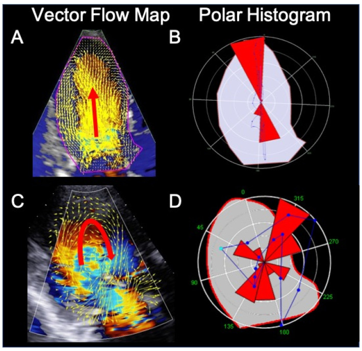
Figure 3.
Intracardiac flow analysis of a normal subject (A) and (B) and a patient with dilated cardiomyopathy (C) and (D). In the normal subject, during diastole, left ventricular (LV) filling occur mainly along a longitudinal axis (panel (A), red arrow). Panel (B) shows the intensity-weighted polar histogram representing the distribution and intensity of the LV hemodynamic forces occurring during the entire heartbeat. The hemodynamic forces (in red) are aligned along the LV base–apex direction according with the normal emptying–filling process of the LV. In the cardiomyopathy patient, LV filling is abnormal, with flow circulating along the posterolateral wall and rotating anteriorly at the level of the left ventricular apex (panel (C), red arrow). The intensity-weighted polar histogram (panel (D)) shows a dispersed distribution of the intraventricular hemodynamic forces. Images were obtained using the HyperDoppler software of an Esaote Mylab X8 echo-scanner without contrast injection.

