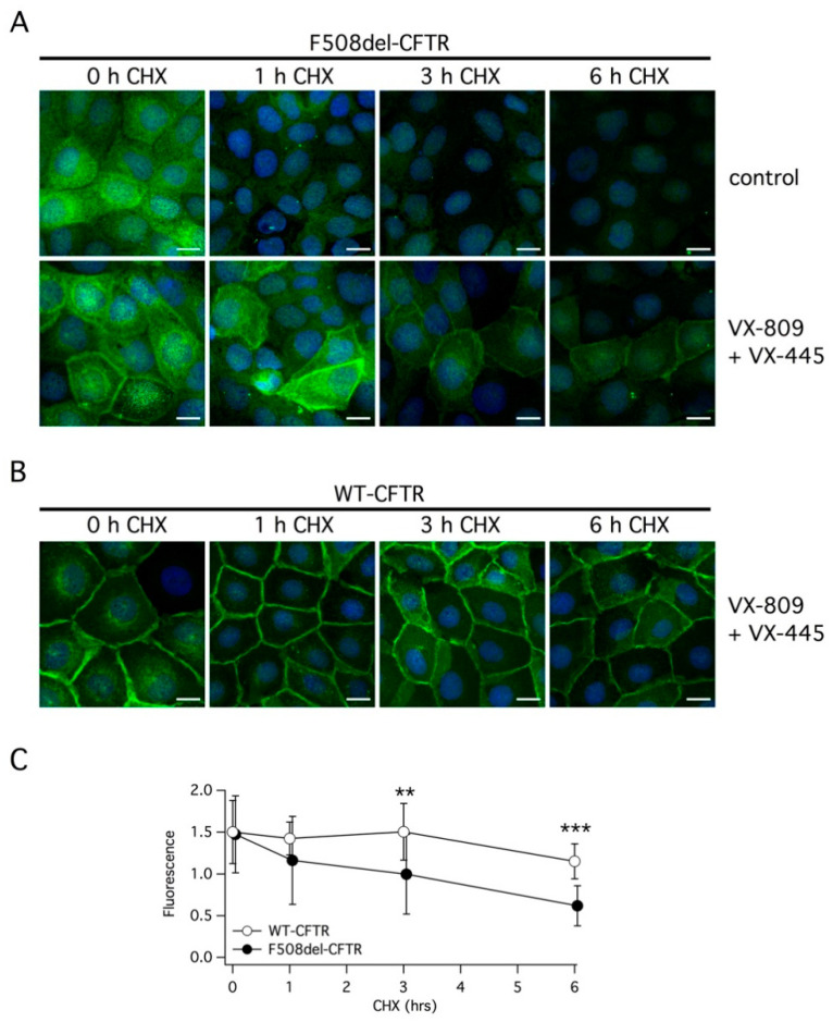
Figure 7.
Analysis of CFTR subcellular localization. (A,B) Representative images showing the detection of F508del-CFTR or wild type CFTR protein in CFBE41o- cells by immunofluorescence. Cells were incubated with vehicle (DMSO) or with the corrector combination (5 µM VX-445 plus 1 µM VX-809) for 24 h. Cells were immediately fixed or treated for the indicated time (1–6 h) with CHX and then fixed (scale bar: 15 µm). (C) Analysis of CFTR protein expression in the plasma membrane at different times following CHX addition. **, p < 0.01; ***, p < 0.001.

