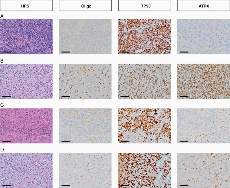
Figure 1.
Histological characteristics of adult diffuse hemispheric gliomas, H3 G34-mutant (Scale bar = 100 µm). (A) PNET-like morphology, characterized by small round cells, with anaplastic features including endothelial cell proliferation, negative Olig2 immunostaining, positive TP53 staining and ATRX loss. (B) Undifferenciated glial morphology, characterized by irregularly shaped nuclei and poorly delimited cytoplasms with anaplastic features including endothelial cell proliferation, positive Olig2 (reactive glial cells), ATRX, and TP53 stainings. (C) Monstrocellular morphology, characterized by multinucleated cells, with anaplastic features including endothelial cell proliferation, necrosis, negative Olig2 immunostaining, positive TP53 staining, and ATRX loss. (D) Oligoid morphology, characterized by small cells with a clear peri-nuclear halo, with anaplastic features including endothelial cell proliferation and 2 mitoses, positive Olig2 (reactive glial cells) and TP53 immunostaining and ATRX loss.

