Figure 2.
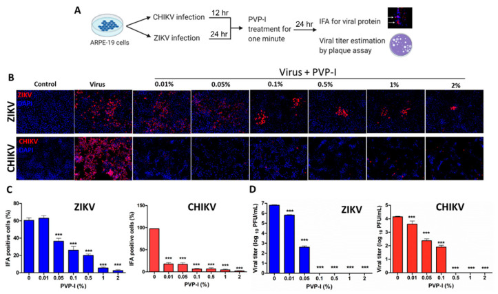
PVP-I exposure attenuates viral replication and production of viral progeny in retinal pigment epithelial cells. Schematic representation of the experimental design (A). ARPE-19 cells were infected with ZIKV and CHIKV for 24 h and 12 h, respectively. Infected cells were treated with indicated concentrations of PVP-I for one minute and extensively rinsed to remove residual PVP-I, and cultured for another 24 h in fresh media. Viral replication was assessed by immunofluorescent detection of viral antigens (red) using anti-Flavivirus 4G2 (ZIKV) and anti-CHKV E1 antibodies in fixed cells. The cell nuclei were counterstained using DAPI (blue). The images were captured at 20X magnification using the Keyence BZ-X800 series microscope (B). The cells expressing viral protein were counted and presented as IFA-positive cells (%) relative to the total number of cells from four independent fields (C). The culture supernatant was used to perform plaque assay on the Vero cell monolayer and the viral titer was expressed as log10 PFU/mL (D). One-way ANOVA with Dunnett’s test was used for statistical analysis, wherein *** p < 0.001.

