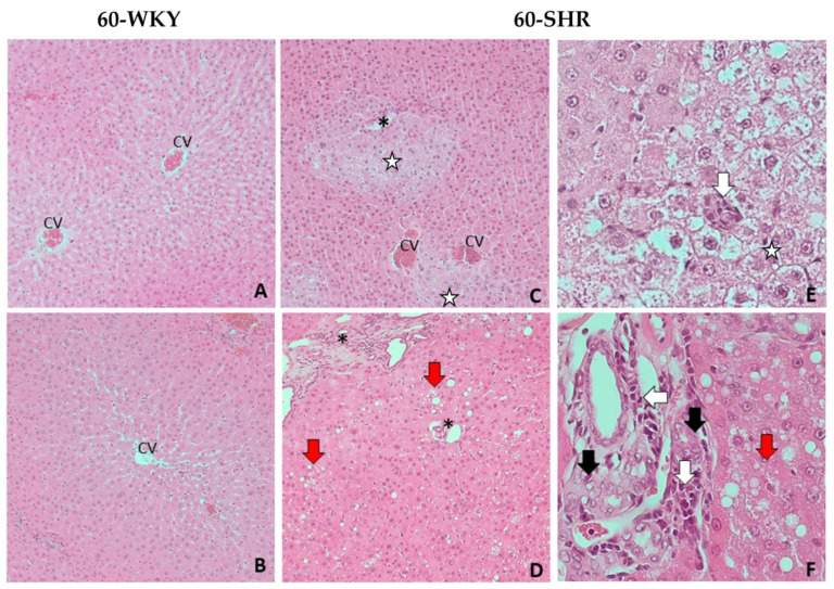
Figure 3.
Histopathological picture of livers of 60-week-old rats. Picture shows the results of histopathological examination of the following groups: 60-week-old Wistar Kyoto rats (60-WKY) and 60-week-old Spontaneously Hypertensive Rats (60-SHR). (A–D)—liver parenchyma at magnification of the lens 10×; (E)—hepatocytes with hydrophic degeneration at magnification of the lens 40×; (F)—portal fields at magnification of the lens 40×. CV—central vein; *—portal field; white arrow—foci of mononuclear cell infiltration; black arrow—cholangiocyte hyperplasia; red arrow—steatosis foci; white star—foci of hydrophic degeneration.

