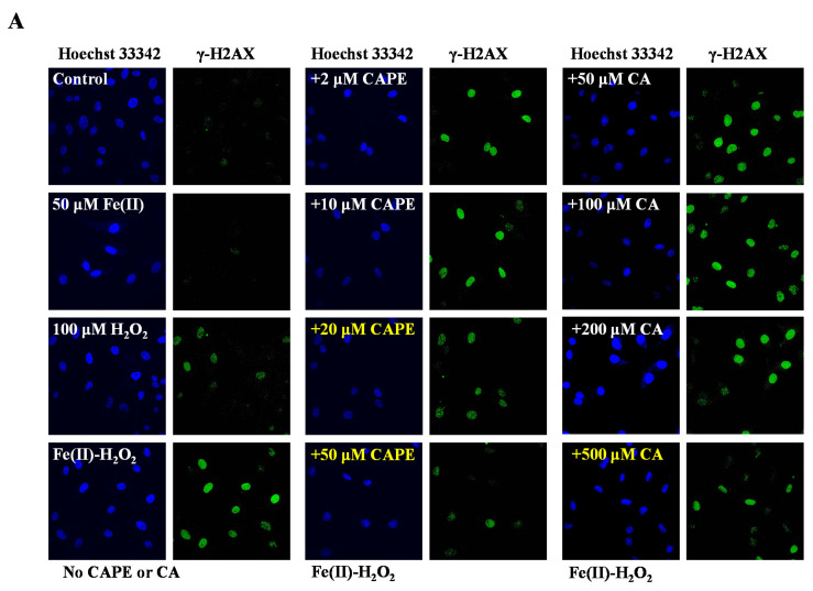
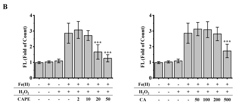
Figure 2.
Inhibition of CAPE and its analogues on the formation of γ-H2AX induced by iron-overload in HeLa cells, as measured by immunofluorescence staining. (A) Representative images of HeLa cells stained for Hoechst 33342 (nuclear stain) monoclonal antibodies of γ-H2AX under different conditions as indicated. (B) Quantitation of γ-H2AX formation in HeLa cells under different conditions as indicated. *** Significant difference from the Fe(II)–H2O2 groups, p < 0.001. The experiment was repeated three times, and thirty cells were randomly selected in different microscopic fields for statistical analysis in each group.


